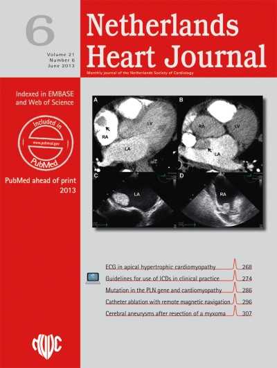
A 63-year-old previously healthy man presented with deep vein thrombosis and dyspnoea. He developed atrial fibrillation during hospitalisation. A CT scan of the chest revealed filling defects of the atria and ventricles (Fig. 1, panel a and b). Upon transoesophageal echocardiography (TEE) (Fig. 1, c–f) intracardiac masses suspect for thrombi were seen in the left atrium (LA) and right atrium (RA). There was no atrial septum defect or patent foramen ovale. The patient was treated with intravenous heparin and a vitamin K antagonist. At follow-up TEE the intracardiac masses disappeared. Despite adequate anticoagulation, the patient developed an intracerebral infarction and died from recurrent aspiration pneumonia. Simultaneous occurrence of massive thrombus formation in both right and left heart chambers is extremely rare [1]. The findings suggest the presence of an intensely activated coagulation, as for instance occasionally seen in patients with systemic inflammatory diseases or malignancies [2]. In the present case, during the short clinical course, none of the common causes of strongly activated coagulation were found to be present.
Fig. 1
CT scan (panel a and b) and TEE images of the intracardiac mass (panel c–f)